sensory nerve impulses from the more than one million
ganglion cells of the retina toward the visual centres in the
brain. The vast majority of optic nerve fibres convey information
regarding central vision.
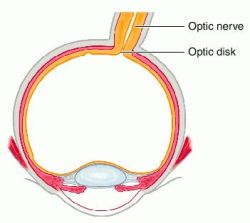
The optic nerve begins at the optic disk at the back
of the eye. The optic disk forms from the convergence of
ganglion cell output fibres (called axons) as they pass out
of the eye. When the nerve emerges from the back of the
eye, it passes through the remainder of the posterior orbit
(eye socket) and through the bony optic canal to emerge
intracranially on the underside of the front of the brain.
At this point the optic nerve from each eye comes together
and forms an X-shaped structure called the optic chiasm.
Here, approximately one-half of the nerve fibres from
each eye continue on the same side of the brain, and the
remaining nerve fibres cross over at the chiasm to join fibres from the opposite eye on the other side of the brain.
This arrangement is essential for producing binocular
vision. Posterior to the optic chiasm, the nerve fibres
travel in optic tracts to various portions of the brain—predominantly
the lateral geniculate nuclei. Fibres from the
lateral geniculate nuclei form the optic radiations that
course toward the visual cortex located in the occipital
lobes in the back of the brain. Some nerve fibres leave the
optic tract without entering the lateral geniculate nuclei
and instead enter the brain stem to provide information
that ultimately determines pupil size.
The retina, optic disk, optic nerve, optic chiasm,
optic tracts, optic radiations, and visual centres of the
brain are topographically organized to correspond to particular
areas of the visual field. Therefore, damage to, or
pressure on, particular portions of these structures can
produce characteristic deficits in a person’s visual field.
The affected person may or may not notice these visual
field defects.
Hiç yorum yok:
Yorum Gönder