It is vitally important that the front surface of the eyeball,
the cornea, remain moist. This is achieved by the eyelids,
which during waking hours sweep the secretions of the
lacrimal apparatus and other glands over the surface at
regular intervals and which during sleep cover the eyes
and prevent evaporation. Maintaining moisture levels is
vital for the normal functioning of the conjunctiva and
cornea. The conjunctiva is the mucous membrane that
lines the eyelid and covers the visible portion of the eyeball
except the cornea.
31 Ağustos 2010 Salı
Orbit
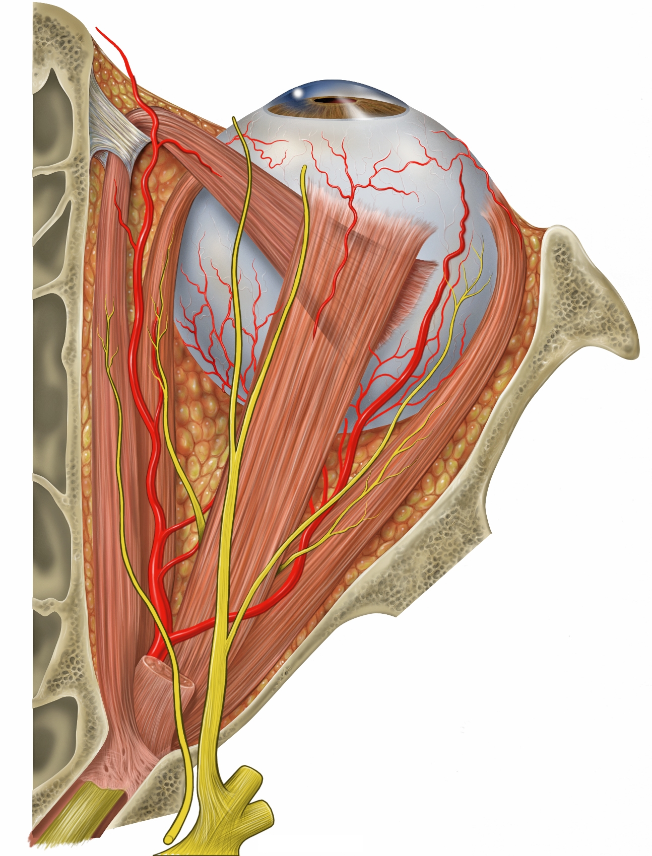
The eye is protected from mechanical injury by being
enclosed in a socket, or orbit, which is made up of portions
of several of the bones of the skull to form a four-sided
pyramid the apex of which points back into the head.
Thus, the floor of the orbit is made up of parts of the maxilla,
zygomatic, and palatine bones, while the roof is made
up of the orbital plate of the frontal bone and, behind this,
by the lesser wing of the sphenoid. The optic foramen, the opening through which the optic nerve runs back into the
brain and the large ophthalmic artery enters the orbit, is
at the nasal side of the apex; the superior orbital fissure is
a larger hole through which pass large veins and nerves.
These nerves may carry nonvisual sensory messages—e.g.,
pain—or they may be motor nerves controlling the muscles
of the eye. There are other fissures and canals
transmitting nerves and blood vessels. The eyeball and its
functional muscles are surrounded by a layer of orbital fat
that acts much like a cushion, permitting a smooth rotation
of the eyeball about a virtually fixed point, the centre
of rotation. The protrusion of the eyeballs—proptosis—
in exophthalmic goitre is caused by the collection of
fluid in the orbital fatty tissue.
Optic Nerve
The optic nerve is the second cranial nerve, which carries
sensory nerve impulses from the more than one million
ganglion cells of the retina toward the visual centres in the
brain. The vast majority of optic nerve fibres convey information
regarding central vision.
The optic nerve begins at the optic disk at the back
of the eye. The optic disk forms from the convergence of
ganglion cell output fibres (called axons) as they pass out
of the eye. When the nerve emerges from the back of the
eye, it passes through the remainder of the posterior orbit
(eye socket) and through the bony optic canal to emerge
intracranially on the underside of the front of the brain.
At this point the optic nerve from each eye comes together
and forms an X-shaped structure called the optic chiasm.
Here, approximately one-half of the nerve fibres from
each eye continue on the same side of the brain, and the
remaining nerve fibres cross over at the chiasm to join fibres from the opposite eye on the other side of the brain.
Optic Disk
The optic disk (also known as the optic nerve head) is a
small region within the retina in which there are no photoreceptors
(i.e., rods or cones); thus there is no image
detection in this area. The optic disk corresponds to the
blind spot in the visual field of each eye. The blind spot of
the right eye is located to the right of the centre of vision
and vice versa in the left eye. With both eyes open, the blind spots are not perceived because the visual fields
of the two eyes overlap. Indeed, even with one eye closed,
the blind spot can be difficult to detect subjectively
because of the ability of the brain to “fill in” or ignore the
missing portion of the image.
Macula Lutea
The macula lutea is the small yellowish area of the retina
near the optic disk that provides central vision. When the
gaze is fixed on any object, the centre of the macula,
the centre of the lens, and the object are in a straight
line. In the centre of the macula is the cone-containing
fovea. Toward the centre of the macula there are no
blood vessels to interfere with vision; thus, in this area of
the retina, vision in bright light and colour perception
are keenest.
Age-related macular degeneration (ARMD) is a relatively
Cones
Cones are the second type of photoreceptor occurring in
the retina of the human eye. They are conical in shape and
are associated with colour vision and perception of fine
detail. Shorter and far fewer than the eye’s rods, cones are
less sensitive to low illumination levels and are mediators
of photopic rather than scotopic (Greek skotos, “dark”)
vision. Cones are mostly concentrated within the central
retina (macula), which contains the fovea (depression in
the retina), where no rods are present. In contrast, the
outer edges of the retina contain few cones and many rods.
Chemical changes that occur when light strikes the cones are ultimately relayed as impulses to optic nerve fibres
that enter the brain.
The Retina
The retina is a layer of nervous tissue that covers the inside
of the back two-thirds of the eyeball, in which stimulation
by light occurs, initiating the sensation of vision. The retina
is actually an extension of the brain, formed
embryonically from neural tissue and connected to the
brain proper by the optic nerve. The retina functions specifically
to receive light and to convert it into chemical
energy. The chemical energy activates nerves that conduct
the electrical messages out of the eye into the higher
regions of the brain.
Pigmented Epithelium
Separating the choroid (the middle tunic of the globe, or
eyeball) from the retina proper is a layer of pigmented
cells, known as the pigment epithelium. This layer of the
retina acts as a restraining barrier to the indiscriminate
diffusion of material from the blood in the choroid to the
retina. The retina ends at the ora serrata, where the ciliary
body begins. The pigment epithelium continues forward
as a pigmented layer of cells covering the ciliary body; farther
forward still, the epithelium covers the posterior
surface of the iris and provides the cells that constitute
the dilator muscle of this diaphragm. Next to the pigment
epithelium of the retina is the neuroepithelium, or rods
and cones. Their continuation forward is represented by a
second layer of epithelial cells covering the ciliary body;
the term ciliary epithelium is used to describe the two layers
of cells that are the embryological equivalent of the
retinal pigment epithelium and the receptor layer (rods
and cones) of the retina. The unpigmented layer of the
ciliary epithelium is continued forward over the back of
the iris, where it acquires pigment and is called the posterior
iris epithelium.
Dilator Muscle
The dilator muscle of the iris involuntarily contracts as
available light decreases, thus dilating the pupil. Pupillary
dilation is controlled primarily by the sympathetic nervous
system. Interruption of the innervation of the dilator
muscle can cause an abnormally small pupil, a condition
seen as part of Horner syndrome. Traumatic rupture of
iris muscles can cause an irregularly shaped pupil. Dilator
muscles can also be found in other parts of the body such
as the nose, where the dilator naris muscle aids in widening
the nostrils.
Pupil
Light must pass through the pupil before it can reach
the lens and be focused onto the retina. Parasympathetic
nerve fibres from the third (oculomotor) cranial nerve
innervate the muscle of the iris that causes constriction
of the pupil, whereas sympathetic nerve fibres control
dilation. The pupillary aperture also narrows when
focusing on close objects and dilates for more distant
viewing. At its maximum contraction, the adult pupil
may be less than 1 mm (0.04 inch) in diameter, and
it may increase up to 10 times to its maximum diameter.
The size of the human pupil may also vary as a result of
age, disease, trauma, or other abnormalities within the
visual system, including dysfunction of the pathways
controlling pupillary movement. Thus, careful evaluation
of the pupils is an important part of both eye and
neurologic exams.
Iris
The most anterior portion of the uvea is the iris. This is
the only portion that is visible to superficial inspection,
appearing as a perforated disc, the central perforation, or
pupil, varying in size according to the surrounding illumination
and other factors. A prominent feature is the
collarette at the inner edge, representing the place of
attachment of the embryonic pupillary membrane that, in
embryonic life, covers the pupil. As with the ciliary body,
with which it is anatomically continuous, the iris consists
of several layers: namely, an anterior layer of endothelium,
the stroma; and the posterior iris epithelium. The stroma
contains the blood vessels and the two sheets of smoothmuscle, the sphincter and dilator muscles, that control the
contraction (constriction) and the expansion (dilation) of
the iris, respectively. In addition, the stroma contains pigment
cells that determine the colour of the eye.
Uveal Tract
The middle coat of the eye is called the uvea (from the
Latin for “grape”) because the eye looks like a reddish-blue
grape when the outer coat has been dissected away. The
posterior part of the uvea, the choroid, is essentially a
layer of blood vessels and connective tissue sandwiched
between the sclera and the retina. The forward portion of
the uvea, the ciliary body and iris, is more complex, containing
as it does the ciliary muscle and the sphincter and
dilator of the pupil.
The blood supply responsible for nourishing the retina
consists of the retinal and uveal circulations, both of which
derive from branches of the ophthalmic artery. The two
systems of blood vessels differ in that the retinal vessels,
which supply nutrition to the innermost layers of the retina,
derive from a branch of the ophthalmic artery, called
the central artery of the retina, that enters the eye
with the optic nerve, while the uveal circulation, which
supplies the middle and outer layers of the retina as well as
the uvea, is derived from branches of the ophthalmic artery
that penetrate the globe independently of the optic nerve.
Cornea
The cornea is the transparent window of the eye. It is
about 12 mm (0.5 inch) in diameter and, except at its margins,
contains no blood vessels. However, it does contain
many nerves and is very sensitive to pain or touch. It is
nourished and provided with oxygen anteriorly by tears
and is bathed posteriorly by aqueous humour. It protects
the pupil, the iris, and the inside of the eye from penetration
by foreign bodies and is the first and most powerful
element in the eye’s focusing system. As light passes
through the cornea, it is partially refracted before reaching
the lens. The curvature of the cornea, which is spherical
in infancy but changes with age, gives it its focusing power;
when the curve becomes irregular, it causes a focusing
defect called astigmatism, in which images appear elongated
or distorted.
The eye
The eyeball can be viewed as the fusing together of a small
portion of a small, strongly curved sphere with a large portion
of a large, not so strongly curved sphere. The small
piece, occupying about one-sixth of the whole, has a radius
of 8 mm (0.3 inch); it is transparent and is called the cornea;
the remainder, called the scleral segment, is opaque
and has a radius of 12 mm (0.5 inch). The ring where the
two areas join is called the limbus. Thus, on looking
directly into the eye from in front one sees the white sclera
surrounding the cornea; because the latter is transparent
one sees, instead of the cornea, a ring of tissue lying within the eye, the iris.
The iris is the structure that determines
the colour of the eye.
Kaydol:
Yorumlar (Atom)